4D NMR Structure
Discover the 3D structure of novel protein targets with our cutting-edge NMR service, designed to streamline your drug discovery journey. In just 3-4 weeks, our unique 4D-NMR protocol delivers comprehensive target domain structures, while our tailored services address additional NMR requirements.
Leveraging our 4D-GRAPHS algorithm, we measure and analyze two 4D NOESY spectra from a single 13C, 15N labeled protein sample, ensuring accurate and complete assignments of sidechain and backbone resonances. This information then guides the automated assignment of long-range NOEs and structure refinement using autoNOE-Rosetta.
Why to choose AI|ffinity’s 4D NMR-based method for protein structure determination?
Our new ML-based version, 4D-GRAPHS, enables automatic, all protein atom resonance assignment using only two 4D NMR spectra, reducing measurement time. For small proteins, a single 4D spectrum is sufficient. These resonance assignments enable 3D structure determination using standard tools.
Advantages:
- Suitable for flexible proteins: if they are soluble and stable at concentrations as low as 0.1-0.2 mM, with the typical range being 0.2 mM to 1 mM; non need for crystallization.
- Speed and Cost-Effectiveness: up to 6x faster and 3x cheaper than X-ray crystallography and up to 2x faster than standard 3D NMR methods.
- Handling Larger Proteins: thanks to the enhanced 4D peak resolution.
- Less Human Input: streamlined spectrum analysis and structure calculations reduce human intervention and related errors.
- Market-Leading Speed: only one 4D spectrum is needed for follow-up structures, such as mutants or protein-ligand complexes.
- Insight Into Protein Dynamics: and mechanisms of action.
- Mapping Interactions: with other molecules and is ideal for weak binders (hit compounds), offering a competitive edge.
- Natural Environment Representation: unlike X-ray crystallography and Cryo-EM that provide structural information about proteins in an artificially "frozen" state.
- No Installation Needed: the algorithm and all necessary software for structure calculations will be provided as a cloud services, including technical support.
Disadvantages:
- Protein Size Limit: at ~30 kDa, although this limit is expected to increase with new Ultra-high field NMR spectrometers (1.2 GHz).
- Dependence on Third-Party Software: for raw spectra processing, which can make the pipeline more complex, though this issue is common to all NMR software for resonance assignment.
- Requirement for High Protein Concentrations: typically 0.5 mM to 2 mM for globular proteins, with larger ones often requiring the upper end for adequate signal-to-noise ratios, risking potential aggregation or precipitation.
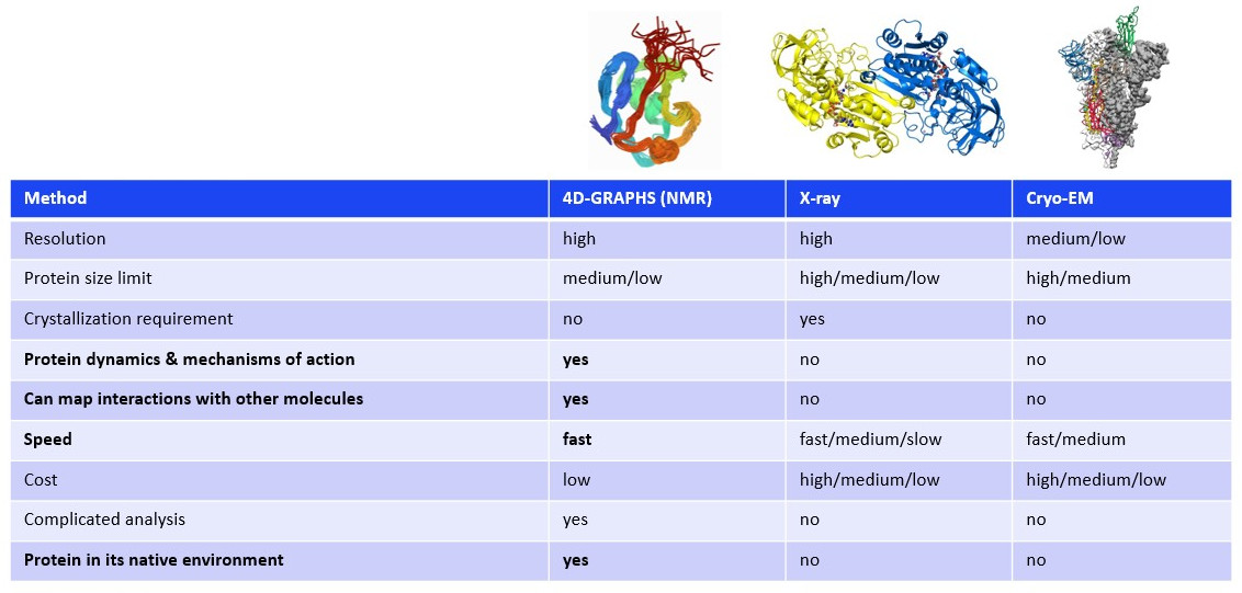
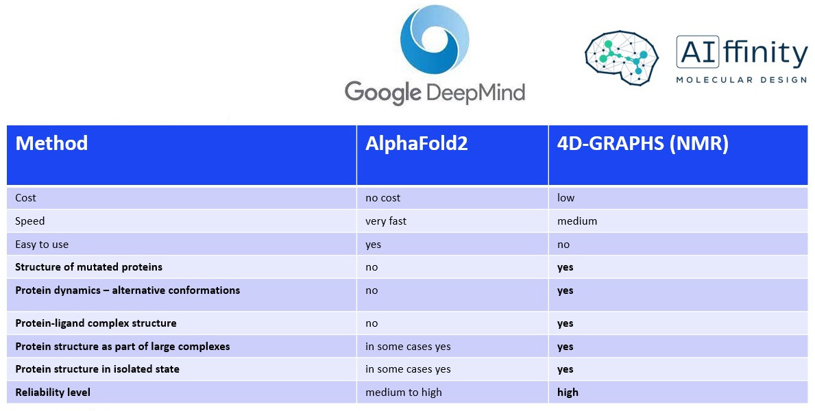
What are the comparative strengths and weaknesses of AI|ffinity's 4D NMR-based technique for protein structure determination versus other available experimental and computational methods?
In addition to 4D NMR structure determination, we also perform a comprehensive array of standard NMR experiments within our drug discovery projects.
Ligand-observe methods (1D NMR): focus on the ligand's spectral properties and have no target size limitations.
- binding validation
- ligand epitope mapping
- ligand epitope mapping
- binding constand (Kd) determination
- competition assays
Target-observe methods (2D NMR): focus on the protein's spectral properties and do have size limitations (~30 kDa).
- mapping of interactions with the protein
- protein conformational changes induced by ligand binding
Leveraging our 4D-NMR technology and computer-aided drug design (CADD), we're adept at designing potent inhibitors for biological targets. Trust AI|ffinity to accelerate your drug discovery process.
References:
- Evangelidis T, et al., "Automated NMR resonance assignments and structure determination using a minimal set of 4D spectra”. Nat Commun. 2018 Jan 26;9(1):384.
- Marcos E, et al., "De novo design of a non-local β-sheet protein with high stability and accuracy". Nat Struct Mol Biol. 2018 Oct 29.
- Hanáková K, et al., "Comparative phosphorylation map of Dishevelled 3 links phospho-signatures to biological outputs". Cell Commun Signal. 2019 Dec 23;17(1):170.
- Harnoš J, et al., "Dishevelled-3 conformation dynamics analyzed by FRET-based biosensors reveals a key role of casein kinase 1". Nat Commun. 2019 Apr 18;10(1):1804.
- Yperman K, et al., "Distinct EH domains of the endocytic TPLATE complex confer lipid and protein binding". Nat Commun. 2021 May 24;12(1):3050.
- De Vos T, et al., "Structural basis for the mechanism and antagonism of receptor signaling mediated by Interleukin-9 (IL-9)". bioRxiv 2022.12.30.522308.